You must be signed in to read the rest of this article.
Registration on CDEWorld is free. You may also login to CDEWorld with your DentalAegis.com account.
Cinnamon flavoring agents (eg, cinnamic aldehyde, cinnamic acid, and cinnamon oil) are commonly added to oral hygiene products, foods, chewing gum, and other products to give them a pleasant flavor. However, cinnamon flavoring agents are known to act on the skin and the mucosa as irritants or sensitizers.1-6 Although contact stomatitis is not a widespread phenomenon, cinnamon has been reported to be a cause, but the symptoms attributed to cinnamon are unknown in many cases.1,3,7
Various products contain cinnamon flavoring agents, and most of the reported contact stomatitis cases are caused by toothpaste or chewing gum.2,4,5,8-15 It is said that the clinical signs and symptoms of these cases are nonspecific, though the signs tend to be present at sites that are in direct contact with the causative agents. Contact stomatitis caused by toothpaste has been reported to induce signs and symptoms in the lips,2,4,5,9,11-13 gingiva,4,5,9,11,12 tongue,9,11,13 and perioral skin.4,5,11,13 Reported clinical features include erythema,4,9,11,12 swelling,2,9,11-13 desquamation,11 peeling,2 epithelial sloughing,12 ulceration,9,11-13 glossitis,9 and cheilitis.2-5,9,11 Contact stomatitis caused by chewing gum has been reported to exhibit white lesions,8,9,14,15 peeling,10 epithelial sloughing,8 and erythema10 in the buccal mucosa,8,10,14 the tongue,8,10,15 and the gingiva.8
To treat contact stomatitis apparently caused by cinnamon, it is important to discontinue the intake of cinnamon flavoring agents. It is necessary to find the possible causes of the disease by understanding the characteristic clinical features and conducting careful patient interviews.
Materials and Methods
The authors examined the records from the database of the Stomatology Center at Baylor College of Dentistry in Dallas, Texas, and identified 65 cases from 1985 through 1998 that were classified as contact stomatitis caused by cinnamon flavoring agents. In 37 of the 65 cases, causative agents were identified, and the signs and symptoms disappeared after the patients discontinued the use of these agents. These 37 cases were used for this study. The other 28 cases were excluded because of the absence of records confirming the disappearance of lesions.
The records examined contained information such as the types of oral hygiene products being used by the patients and the frequency of gum chewing. Patients kept food diaries for 1 to 2 weeks, and the contents of their diets were evaluated if necessary. Frequently used flavoring agents were particularly evaluated. In some cases, evaluation included patch tests and biopsies. This study analyzed the descriptions of clinical features, sites of onset, and types of causative agents from each case. The results of the patch tests for cinnamon, histopathological examinations, and the treatment for each patient were also analyzed.
Results
The results of the 37 cases are shown in Table 1. There were 5 men and 32 women with an average age of 48.4 ± 15.2 years, ranging from ages 20 to 80, at the time of presentation. Patients had been afflicted with contact stomatitis for 1 month to 8 years before the diagnosis was determined and treatment rendered. Causative agents were classified into 3 categories: toothpaste, chewing gum, and foods. In this study, we regarded beverages as foods. Toothpaste was the largest category (27 cases, 73%), followed by chewing gum (8 cases, 22%) and foods (8 cases, 22%). In 5 cases (14%), patients fit into 2 or more categories. In all of the cases in which toothpaste was the cause, the toothpaste was a tartar-control product. Most of these patients reported brushing their teeth 3 times a day.
Patient interviews in cases caused by contact with chewing gum said that they were “chewing gum for 8 hours a day only on the left side” (Case 19), “chewing 4 to 6 packs of gum every day” (Case 32), or “chewing gum every day since she gave up smoking” (Case 37). Of the 37 cases, 18 (49%) had a histopathological examination of the gingiva or the buccal mucosa. Patch tests for cinnamon were conducted in 15 cases (41%). Both histopathological examinations and patch tests were conducted in 7 cases (19%).
The site where clinical signs and symptoms were most frequently found was the gingiva (26 cases, 70%), followed by the buccal or labial mucosa (16 cases, 39%), the tongue (13 cases, 35%), and the lips (8 cases, 22%) (Table 2). One patient (Case 3) reported a burning sensation as a symptom, but no description of the site of onset was recorded. The clinical features described were erythema (25 cases, 68%), epithelial sloughing (9 cases, 24%), white lesions (8 cases, 22%), lip swelling (6 cases, 16%), edema (6 cases, 16%), fissured tongue (5 cases, 14%), ulceration (4 cases, 11%), granular surface of gingiva (3 cases, 8%), geographic tongue (2 cases, 5%), and other (4 cases, 11%) (Table 2).
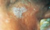
Most of the erythema was observed in the attached gingiva. Twenty cases (54%) included a description of diffuse or generalized erythema of the attached gingiva (Figure 1). Fifteen of these cases were caused by toothpaste. A “velvet-like appearance of gingiva” was a symptom in Cases 4, 6, and 33. Case 27 contained the description “fiery red gingiva.” In 17 cases, gingival erythema was observed in the anterior facial area. Of these, 6 cases (Cases 4, 18, 21, 25, 27, and 35) also showed erythema in the labial mucosa that was in contact with the inflamed facial gingiva. Four cases (Cases 24, 30, 31, and 37) showed gingival erythema in the palatal area.
Epithelial sloughing was noted in the gingiva (Cases 4, 6, 14, 20, 21, 27, and 30), buccal mucosa (Case 11), labial mucosa (Case 18), and tongue (Case 18). Seven of these cases were caused by toothpaste, and the remaining 2 cases were caused by foods or foods plus chewing gum.
In Cases 1, 2, 11, and 13, white lesions or white plaque were found in the buccal mucosa of 1 side (Figure 2). Cases 15 and 26 had white patches or white plaque in the lips. Cases 8, 19, and 26 listed chewing gum as a cause, and white plaque was found in the lateral border of the tongue and the buccal mucosa (Cases 8 and 19) or the lingual gingiva of the same side (Case 26).
Patients in 16 cases (43%) described their symptoms as “sore mouth,” “irritation,” or “burning sensation.” Six of the 8 patients with lesions associated with foods complained of similar symptoms.
The results of the histopathological examination are shown in Table 3. Of the 18 patients who had histopathological examinations, 13 (72%) were diagnosed as having chronic mucositis or chronic gingivitis. Three others showed chronic psoriasiform mucositis. In the superficial lamina propria, inflammatory cell infiltration of plasma cells (8 cases), lymphocytes (5 cases), lymphocytes and histiocytes (4 cases), and eosinophils (2 cases) were recognized. Lichenoid band-like infiltration was observed in 1 case. In the deep layer of the lamina propria, perivascular infiltration was observed in 4 cases. In addition, focally granulomatous inflammation was observed in 4 cases.
Patch tests to determine allergic reactions were performed in 15 patients using standard skin tests for cinnamon compounds, which included 5% cinnamic acid and 2% trans cinnamic aldehyde. Patch tests in 2 cases were conducted for the flavoring agents in the patients’ toothpastes. These agents were later identified as cinnamic aldehyde. Twelve of the 15 patients (80%) showed positive reactions.
Signs and symptoms disappeared in 26 patients (70%) after they stopped using causative agents containing cinnamon. In addition to the discontinuation of causative agents, a topical corticosteroid (0.05% fluocinonide) was prescribed in 11 cases (30%) for the benefit of expediting healing. All 37 patients experienced complete remission without recurrence during 6 months in which they were followed.
Discussion
The cinnamon-containing substance that caused contact stomatitis in most cases was toothpaste. Tartar-control toothpastes usually contain tetrasodium or tetrapotassium pyrophosphates as their anticalculus component.16 Because pyrophosphates have a strong, bitter taste, large quantities of cinnamon flavoring agents are needed in tartar-control toothpastes to mask that taste.16 Cinnamon-induced contact stomatitis caused by toothpaste occurred in various sites, but the gingiva was the most common. Diffuse or generalized erythema was found in the attached gingiva. This characteristic was seen in 20 cases (54%); most were caused by toothpaste. The facial and the palatal/lingual gingiva was affected, but the anterior facial changes were more pronounced. Erythema was described as a “velvet-like appearance of the gingiva” or “fiery red gingiva.” These clinical features were similar to those previously reported as plasma cell gingivitis.17,18 Epithelial sloughing was also most frequently noted in the gingiva. Epithelial sloughing is the most common irritant effect associated with toothpastes and mouthrinses.19 Toothpaste was the cause for 7 of the 9 patients with epithelial sloughing in this study.
Rees has stated that clinical symptoms attributed to toothpaste probably occur most frequently in the gingiva because causative agents contact the gingiva more intensely than other sites during tooth brushing.19 This study included 6 cases of erythema in the labial mucosa in contact with the inflamed facial attached gingiva. The gingiva appears to be the primary site of contact stomatitis caused by toothpaste. The symptoms in the labial mucosa seem to be secondary because the inflammation of the attached gingiva is more widespread and severe than that of the labial mucosa. Other reported clinical signs and symptoms caused by toothpaste, including perioral lesions and angular cheilitis,4,5,11,13 were not observed in this study.
It is characteristic that the symptoms brought on by chewing gum tend to be localized in the buccal mucosa and the tongue compared with those caused by toothpaste.8,10,14,15 There have been reports of leukoplakia in the buccal mucosa14 and squamous cell carcinoma in the lateral border of the tongue15 associated with the use of cinnamon-flavored chewing gum. This study also showed that signs from chewing gum tended to occur in the buccal mucosa and the tongue. Three cases involving chewing gum (Cases 8, 19, and 26) showed white plaque at the lateral border of the tongue and the buccal mucosa of the same side. This phenomenon may have occurred because patients hold gum between the lateral tongue and the buccal mucosa while pausing from chewing.15
Although none of the patients in this study were using hard candies or mints containing cinnamon, they also may cause contact stomatitis. Like chewing gum, candy is associated with lesions localized in the buccal mucosa and the tongue, probably as a result of direct contact. Related lesions have been reported to include erythematous patches, often with associated keratosis or ulceration10 or white plaque.8
Foods containing cinnamon include canned products, curries, Chinese foods, cakes, and biscuits. Cola drinks, carbonated beverages, vermouth and gin also may cause contact stomatitis. Like toothpaste, contact stomatitis caused by foods was accompanied by diffuse or generalized erythema in the attached gingiva, but epithelial sloughing was usually not evident in this study. A sore mouth and a burning sensation are symptoms often seen in contact stomatitis.3,10,11,13 In this study, 16 of the 37 cases had a sore mouth, irritation, or a burning sensation. In particular, most patients having disorders caused by foods characteristically complained of symptoms appearing after meals or after the intake of specific foods or beverages in this study.
It is difficult to identify foods as a cause of contact stomatitis compared with toothpaste or chewing gum. In this study, patch tests or histopathological examinations were conducted in 26 cases. In at least 7 of these patients, exposure to food was a contributing factor (Table 1). The laboratory test most frequently used to aid in the diagnosis of a contact allergy is the patch test.1 Patients are considered to have allergic reactions to cinnamon if their patch test results are positive. They were conducted in 6 of the 8 cases associated with allergy to food substances, and all showed positive reactions to cinnamon.
Dietary analysis was conducted in this group after positive patch testing or suggestive histopathologic findings. Patient maintenance of a food diary is often beneficial in identifying the causes. Because cinnamon is used in a wide variety of foods, patients may be taking cinnamon unknowingly. They were asked to record in their diaries all foods ingested for 1 to 2 weeks (eg, beverages, chewing gum, candies, breath mints, vitamins, or medications). It was also recommended that they record the use and frequency of oral hygiene products (eg, toothpaste, dental floss, mouthrinse, and oral irrigators). Signs and symptoms of contact stomatitis seemed to develop in association with the amount and frequency of intake of the causative agents.5,14,15 In 2 patients (Cases 4 and 32), the problems had been caused by the intake of 3 to 4 bottles per day of a carbonated beverage containing cinnamon. Dark colas are likely to contain this flavoring agent. Foods containing cinnamon were identified from the food diary. The patients discontinued intake and the clinical symptoms disappeared.
Clinical signs of cinnamon-induced contact stomatitis are similar to those of other mucosal diseases such as lichen planus,1,8,10,15 lupus erythematosus,8,10,15 candidosis,10 and early vesiculobullous disease.1 It is necessary to conduct histopathological examinations to exclude these mucosal diseases from the diagnosis. Histopathological examinations were conducted in 18 cases, all of which had nonspecific inflammatory reactions that were not suggestive of a specific mucocutaneous disease.
Few reports have been made on histopathological findings of cinnamon-induced contact stomatitis.1,8-10,14,15 However, these reports exhibit some similarities, though most tissue reactions are nonspecific. Allen and Blozis reported on the histopathological examination of 2 cases that showed a psoriasiform pattern as a characteristic finding.10 On the other hand, Miller and colleagues reported that histopathological examination of 12 cases revealed no psoriasiform changes; rather, the lesions were more characteristic of a lichenoid tissue reaction.8
Histopathological examinations of 18 of the cases in this study revealed chronic psoriasiform mucositis in 3 cases and lichenoid features in only 1. Inflammatory cell infiltration, mainly plasma cells and lymphocytes, was observed in the superficial lamina propria. No evidence of intense plasma cell infiltrate was found in this study, though this has been previously described as a feature of contact allergy to chewing gum.20 Perivascular infiltration in the deep layer appears to be one of the characteristic features of oral contact stomatitis. In particular, Miller and colleagues observed perivascular infiltration in all the cinnamon-induced cases they described.8 This characteristic was also observed in our study (4 of 18 cases). However, it was seen in fewer cases compared with the report by Miller and colleagues.
The existence of focal granulomatous inflammation in the lamina propria was not described in the other papers studying cinnamon-induced contact stomatitis.8,10 It is now commonly believed that histological changes caused by sensitivity to various foods, preservatives, and oral hygiene products may be consistent with the findings of orofacial granulomatosis.21-23 Patton and colleagues have reported on orofacial granulomatosis caused by foods or flavoring intolerance.23 They stated that cinnamic aldehyde is a main provoking factor. Further research is necessary to determine the relationship between orofacial granulomatosis and cinnamon-induced contact stomatitis.
Some of the reactions to cinnamon are allergic.1-3,9,11,12,14 In this study, 15 of the 37 cases had patch tests for cinnamon. Positive reactions were observed in 12 cases (80%). Two common cinnamon flavoring agents are cinnamic aldehyde, the principal component of cinnamon oil, and cinnamic acid. Cinnamic aldehyde is often used in tartar-control toothpastes.16 When cinnamon flavoring agents penetrate the oral mucosa, dendritic cells are activated and sensitization occurs.1 If sensitized patients are exposed to cinnamon flavoring agents again, a delayed hypersensitivity reaction may develop in the sites of contact.1 In other instances, cinnamon-containing products may elicit an irritation contact stomatitis that is difficult to distinguish from an allergic response.5 In some cases, the allergic and irritant effects may overlap. Consequently, it may be important to use patch tests as a part of the diagnostic process.
Definitive diagnosis of contact stomatitis is determined when the discontinuation of the intake of the causes results in the remission of clinical signs and symptoms. This was achieved in all of the 37 cases included in this study. On occasion, the improvement is gradual, and the patient may benefit from the use of a potent topical corticosteroid to suppress the inflammatory reaction, accelerate tissue healing, and to provide symptomatic relief for acute lesions.21,24 A topical corticosteroid was administered to 11 of the patients.
Conclusion
Although contact stomatitis caused by cinnamon-containing substances is relatively uncommon, these 37 cases demonstrate that it may occur with more frequency than previously recognized. The most common origin of the 37 cases was toothpaste; other causes were chewing gum and foods. Although various clinical features were observed, some characteristics were shown. The most commonly affected site was the gingiva, which showed diffuse or generalized erythema and epithelial sloughing. On numerous occasions, patients had been afflicted with contact stomatitis for months to years before the diagnosis was determined and treatment rendered. Identifying the causative agents of such a disorder by understanding characteristic signs and symptoms and conducting an appropriate inquiry is of the utmost importance.
References
1. De Rossi SS, Greenberg MS. Intraoral contact allergy: a literature review and case reports. J Am Dent Assoc. 1998;
129:1435-1441.
2. Maibach HI. Cheilitis: occult allergy to cinnamic aldehyde. Contact Dermatitis. 1986;15:106-107.
3. Drake TE, Maibach HI. Allergic contact dermatiis and stomatitis caused by a cinnamic aldehyde-flavored toothpaste. Arch Dermatol. 1976;112:202-203.
4. Ferlito TA. Tartar-control toothpaste and perioral dermatitis. J Clin Orthod. 1992;26:43-44.
5. Beacham BE, Kurgansky D, Gould WM. Circumoral dermatitis and cheilitis caused by tartar control dentifrices. J Am Acad Dermatol. 1990;22:1029-1032.
6. Fisher AA. Contact stomatitis, glossities, and cheilitis. Otolaryngol Clin North Am. 1974;7:827-843.
7. Pantlin L, Joyston-Bechal S. Sensitivity to flavored toothpaste. Dent Update. 1988;15:425-426.
8. Miller RL, Gould AR, Bernstein ML. Cinnamon-induced stomatitis venenata. Clinical and characteristic histopathologic features. Oral Surg Oral Med Oral Pathol. 1992;73:708-716.
9. Lamey PJ, Lewis MA, Rees TD, et al. Sensitivity reaction to the cinnamonaldehyde component of toothpaste. Br Dent J. 1990;168:115-118.
10. Allen CM, Blozis GG. Oral mucosal reactions to cinnamon-flavored chewing gum. J Am Dent Assoc. 1988;116:664-667.
11. Millard L. Acute contact sensitivity to a new toothpaste. J Dent. 1973;1:168-170.
12. Thyne G, Young DW, Ferguson MM. Contact stomatitis caused by toothpaste. N Z Dent J. 1989;85:124-126.
13. Magnusson B, Wilkinson DS. Cinnamic aldehyde in toothpaste. 1. Clinical aspects and patch tests. Contact Dermatitis. 1975;1:70-76.
14. Mihail RC. Oral leukoplakia caused by cinnamon food allergy. J Otolaryngol. 1992;21:366-367.
15. Westra WH, McMurray JS, Califano J, et al. Squamous cell carcinoma of the tongue associated with cinnamon gum use: a case report. Head Neck. 1998;20:430-433.
16. DeLattre VF. Factors contributing to adverse soft tissue reactions due to the use of tartar control toothpastes: report of a case and literature review. J Periodontol. 1999;70:803-807.
17. Sollecito TP, Greenberg MS. Plasma cell gingivitis. Report of two cases. Oral Surg Oral Med Oral Pathol. 1992;73:690-693.
18. Silverman S Jr, Lozada F. An epilogue to plasma-cell gingivostomatitis (allergic gingivostamatitis). Oral Surg Oral Med Oral Pathol. 1977;43:211-217.
19. Rees TD. Drugs and oral disorders. Periodontology 2000. 1998;18:21-36.
20. Kerr DA, McClatchey KD, Regezi JA. Allergic gingivostomatitis (due to gum chewing). J Periodontol. 1971;42:709-712.
21. Rees TD. Orofacial granulomatosis and related conditions. Periodontology 2000. 1999;21:145-157.
22. Armstrong DK, Biagioni P, Lamey PJ, et al. Contact hypersensitivity in patients with orofacial granulomatosis. Am J Contact Dermat. 1997;8:35-38.
23. Patton DW, Ferguson MM, Forsyth A, et al. Oro-facial granulomatosis: a possible allergic basis. Br J Oral Maxillofac Surg. 1985;23:235-242.
24. Jainkittivong A, Langlais RP. Allergic stomatitis. Semin Dermatol. 1994;13:91-101.
About the Author
Hiroyasu Endo, DDS, PhD
Assistant Professor
Department of Periodontology
Nihon University School of Dentistry at Matsudo
Chiba, Japan
Terry D Rees, DDS, MSD
Professor
Director of Stomatology
Department of Periodontics
Baylor College of Dentistry
Texas A&M University Health Science Center
Dallas, Texas